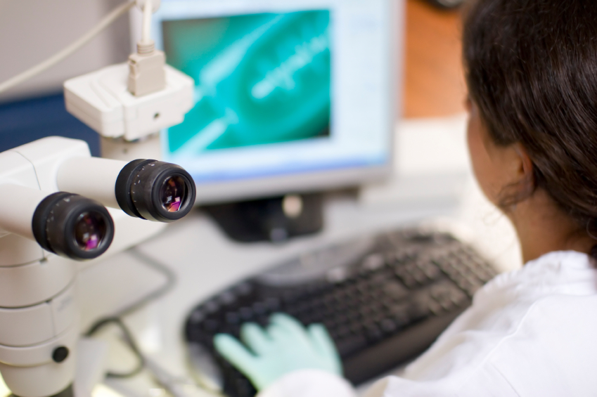
American scientist, Erik Betzig, has just developed a new technique allowing “intracellular processes to be observed in 3D and in real time“.
According to the Sciences & Avenir website, “the main problem with microscopes used to observe objects less than 0.2 micrometres in size is that lasers or rays that substantially damage the sample are needed to study the objects at such resolutions“. Erik Betzig has succeeded in “overcoming this obstacle by improving a technique called ‘light sheet‘”. This involves “taking optical sections of a sample by illuminating it from the side with the famous light sheet obtained using a laser and a lens“. In actual terms, an image can be taken every two milliseconds. This technique allows “objects the size of a molecule or protein to be observed in 3D“.
Scienceetavenir.fr (Joël Ignasse) 27/10/2014

